
 In Pregnancy?")
Image: Shutterstock
Crown-rump length or CRL is the measurement of the fetus from top to bottom. In this post, we explain how to measure crown-rump length in pregnancy.
During pregnancy, your doctor may prescribe several prenatal tests, scans, or procedures to ensure the proper growth and safety of the developing fetus.
The doctor suggests these tests based on genetics, medical history, and other pregnancy-related complications. However, CRL is a vital procedure that helps evaluate the baby’s growth and development. Read on to learn more about CRL, what it tells about the baby, and how it is measured.
What Is CRL?
CRL or crown-rump length is the measurement of the fetus or C-shaped embryo from the top to bottom or the crown to rump, excluding the limbs. Thisultrasound scanis one of the vital measures necessary for antenatal care during thefirst trimesterand is done between six andthirteen weeksapproximately.
The crown to rump length, also known as the greatest length (GL), is an accurate measurement in centimeters to determine the fetal age.It also helps to assess the accurate gestational age, which helps doctors to know more about the baby’s health and also estimate the date of delivery(1)(2).
 Quick fact
What Does CRL Tell You About A Baby’s Health?
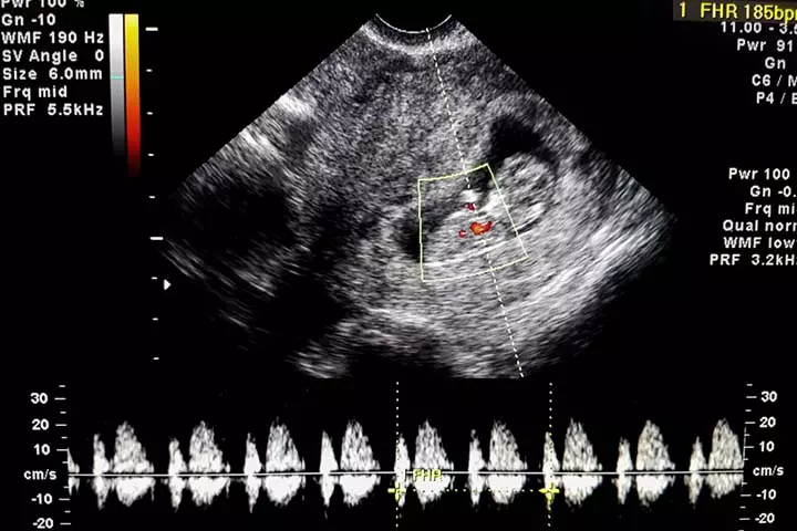
Image: Shutterstock
The CRL scan will let the doctor learn about the early developmental stages of your baby. Some of them include:
- Tracking heart rate:The embryonic heart rate from 6-6.2 weeks is around 100bpm or beats per minute, while it increases to 120-160bpm between 6.3 and 7 weeks(3). Your doctor will keep track of this through the crown-rump length and gestation age to determine how healthy your baby is.
- Identifying complications:The scan helps detect complexities that may arise during the earlystages of pregnancy. For instance, if MSD (mean gestational sac diameter) – CRL = less than 5mm, the risk of pregnancy loss is around 8%.If the difference between MSD and CRL is between 6 and 10mm, the risk is 3-4%.And if it is greater than 10mm, the loss would be less than 1%(4). Identifying these signs in time will help you avoid additional complications.
- Monitoring fetal growth:As the CRL measures the fetal development, any delay or discrepancy in the fetal growth could be a sign of impending loss(5). That is why doctors keep monitoring the length and mass throughout the pregnancy.
- Recognizing abnormalities:Comparing CRL and mean sac diameter helps to detect fetal size and growth retardation. Hence, the ultrasonographic screening during the first trimester also helps the doctor figure out if there are any chromosomal anomalies and risks such asPatau syndromeiXChromosomal condition causing severe physical and intellectual disabilities,Down syndromeiXGenetic condition resulting in distinctive physical and intellectual limitations, andEdwards syndromeiXSevere chromosomal condition affecting fetal growth and causing developmental delays(6).
 Did you know?
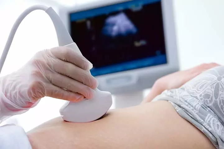
Image: Shutterstock
After the first trimester, CRL might not be an accurate measurement because the baby grows in size and becomes active.And so, some other obstetric ultrasound sonography scans are used to keep a watch on aspects of fetal weight and fetal anatomy such as femur length, abdominal circumference, and fetal biparietaldiameteriXDistance between bones forming the top and sides of the skull, used to estimate the size of the fetus(7).
How Is CRL Measured?
Doctors use various formulations to evaluate the CRL, gestational age, and other aspects. One of them is a simple math equation to find the gestation age using the CRL. According to that, the gestational age equals to six weeks plus crown-rump length multiplied by a number of days [GA = 6 weeks + (CRL x days)] (2).
此外,医生使用crown-rump勒ngth chart as a reference to perform the primary evaluation. An example of such, the Monash chart, is given below(8).
| CRL (mm) | GA (wks, days) | GA (days) |
|---|---|---|
| 1.0 mm | 6W1D | 42.9 |
| 1.5 mm | 6W1D | 43.3 |
| 2.0 mm | 6W2D | 43.7 |
| 2.5 mm | 6W2D | 44.1 |
| 3.0 mm | 6W3D | 44.5 |
| 3.5 mm | 6W3D | 45.0 |
| 4.0 mm | 6W3D | 45.4 |
| 4.5 mm | 6W4D | 45.8 |
| 5.0 mm | 6W4D | 46.2 |
| 5.5 mm | 6W5D | 46.6 |
| 6.0 mm | 6W5D | 47.0 |
| 6.5 mm | 6W5D | 47.4 |
| 7.0 mm | 6W6D | 47.8 |
| 7.5 mm | 6W6D | 48.2 |
| 8.0 mm | 7W0D | 48.6 |
| 8.5 mm | 7W0D | 49.1 |
| 9.0 mm | 7W0D | 49.5 |
| 9.5 mm | 7W1D | 49.9 |
| 10.0 mm | 7W1D | 50.3 |
| 10.5 mm | 7W2D | 50.7 |
| 11.0 mm | 7W2D | 51.1 |
| 11.5 mm | 7W3D | 51.5 |
| 12.0 mm | 7W3D | 51.9 |
| 12.5 mm | 7W3D | 52.3 |
| 13.0 mm | 7W4D | 52.7 |
| 13.5 mm | 7W4D | 53.2 |
| 14.0 mm | 7W5D | 53.6 |
| 14.5 mm | 7W5D | 54.0 |
| 15.0 mm | 7W5D | 54.4 |
| 15.5 mm | 7W6D | 54.8 |
| 16.0 mm | 7W6D | 55.2 |
| 16.5 mm | 8W0D | 55.6 |
| 17.0 mm | 8W0D | 56.0 |
| 17.5 mm | 8W0D | 56.4 |
| 18.0 mm | 8W1D | 56.8 |
| 18.5 mm | 8W1D | 57.3 |
| 19.0 mm | 8W2D | 57.7 |
| 19.5 mm | 8W2D | 58.1 |
| 20.0 mm | 8W2D | 58.5 |
| 20.5 mm | 8W3D | 58.9 |
| 21.0 mm | 8W3D | 59.3 |
| 21.5 mm | 8W4D | 59.7 |
| 22.0 mm | 8W4D | 60.1 |
| 22.5 mm | 8W5D | 60.5 |
| 23.0 mm | 8W5D | 60.9 |
| 23.5 mm | 8W5D | 61.4 |
| 24.0 mm | 8W6D | 61.8 |
| 24.5 mm | 8W6D | 62.2 |
| 25.0 mm | 9W0D | 62.6 |
| 25.5 mm | 9W0D | 63.0 |
| 26.0 mm | 9W0D | 63.4 |
GA:gestational age, W: weeks, and D: days.
The chart is considered accurate for 6 to 9 weeks of the gestation period. However, you should not rely on it completely as it is only an approximation. As every pregnancy is different, you can come across multiple dissimilarities. Therefore, it is always better to consult your doctor and get regular reports regarding the fetal well-being rather than rely on the charts alone.
Did you try estimating your baby’s health using CRL measurements? Let us know about your experiences.
Frequently Asked Questions
1. Does the CRL indicate gender?
Fetal gender can be reliably calculated when the CRL is ≥60 mm (gestational age ≥12+2). Male gender can be determined even when the CRL is ≥55mm. However. if the CRL is less than 50 mm (gestational age (9).
2. Can the CRL measurement be wrong?
The CRL can be incorrectly calculated at times. Inaccuracies by even a margin of 5-10mm may cause significant errors in the fetus’ gestational age calculation. Therefore, to increase the accuracy of the measurements, specialists use these five criteria — accurate magnification, fetus neutrality, the horizontal position of the fetus, precise placement of calipers, and the presence of anamniotic fluidiXA clear fluid surrounding and protecting the fetus in the uterus during pregnancypocket under the fetus’ chin(10).
3. What if CRL is less than expected?
A shorter than average Crown-Rump Length (CRL) during pregnancy can be a sign of an abnormality, but it’s not always the case. It could also mean that the pregnancy is not as advanced as you thought(11).
Crown-rump length in pregnancy is an important procedure used to measure the length of the fetus. The ultrasound scan enables doctors to determine the development of the fetus by assessing its length from the crown to the rump. The scan is done in the early developmental stage and helps doctors detect complications, monitor fetal growth, and track the heart rate. Doctors use the CRL to evaluate gestation age too. They usually refer to a standard chart to find out the gestational age of the fetus; however, since every pregnancy is different, the values of gestational age may differ from the chart. So, it is better to consult a doctor for all your queries rather than depending just on the chart.
Infographic: What Are The Criteria For Crown-Rump Length Measurement?
Practitioners strictly follow the crown-rump measurement criteria to increase the results’ accuracy. Failing to meet the criteria may cause false positive or negative results, which could impact further pregnancy care. Check out the infographic to know how the practitioners get accurate CRL measurements.
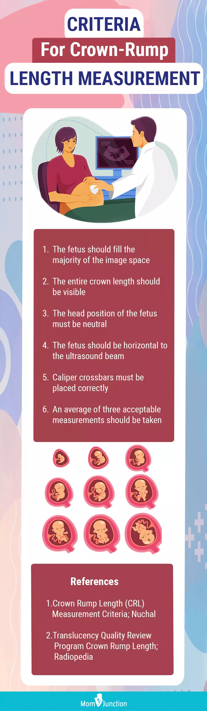
Illustration: Momjunction Design Team
Get high-quality PDF version by clicking below.
 Download Infographic

Key Pointers
- CRL evaluation is carried out in the first trimester to measure the baby’s length and estimate the due date based on the results.
- The evaluation is based on formulas and a crown-rump length chart.
- Besides gestational age, doctors can also evaluate the baby’s heart rate and overall development from the CRL scan.
Discover the precise techniques for measuring nuchal translucency and crown-rump length in a 12-13 week scan. Unveil step-by-step instructions and expert insights to ensure utmost accuracy.
References:
2. D. H. Hareva, I. A. Lazarusli, and Suryasari;Automatic Gestational Age Estimation Based on Crown Rump Length and Gestational Sac; Journal of Image and Graphics (2016)
3. S. K. Rodgers, C. Chang, J. T. DeBardeleben, and M. M. Horrow;Normal and Abnormal US Findings in Early First-Trimester Pregnancy: Review of the Society of Radiologists in Ultrasound 2012 Consensus Panel Recommendations; The Radiological Society of North America (2015)
4. S. Mukherjee;Timing Of Gestational Arrest Prior To Miscarriage; Graduate School of Vanderbilt University (2014)
5. A. Kurjak, S. Kupesic, J. M. Carrera, and B. Ahmed;Ultrasound Evaluation of Abnormal Early Pregnancy; Donald School Journal of Ultrasound in Obstetrics and Gynecology (2008)
6.Down Syndrome; Center of prenatal ultrasonographic diagnostics
7. W. Akhtar et al.;超声胎儿生物统计学为巴基斯坦的一个图表cohort; Eastern Mediterranean Health Journal (2011)
8. P. Delpachitra et al.;Ultrasound Reference Chart Based on IVF Dates to Estimate Gestational Age at 6–9 weeks’ Gestation; ISRN Obstetrics and Gynecology (2012)
9. Marek Lubusky et al;Ultrasound evaluation of fetal gender at 12-14 weeks; National Library Of Medicine (2012)
10. Dominik Jakubowsk et al.;The crown-rump length measurement — ISUOG criteria and clinical practice; Ginekologia Polska (2020)
11. Yin Xu et al.;Understanding how 4 abnormal ultrasound findings relate to miscarriage risk; The University of Texas Southwestern Medical Center.
12.Antenatal Screening for Down Syndrome and other Conditions; National Screening Unit; Government of Newzealand


 In Babies Why It Is Done And Possible Side Effects")


%20In%20Pregnancy?)
%20In%20Pregnancy?)
%20In%20Pregnancy?)